


Imaging Advances in Forensic Anthropology
Forensic anthropology heavily relies on imaging technologies, playing a crucial role in documenting various cases. The progression of digital photography has empowered anthropologists to meticulously capture scenes and skeletal remains. However, standard photos are often insufficient for detailed analysis, prompting the adoption of X-ray scanning technology to assist anthropologists.
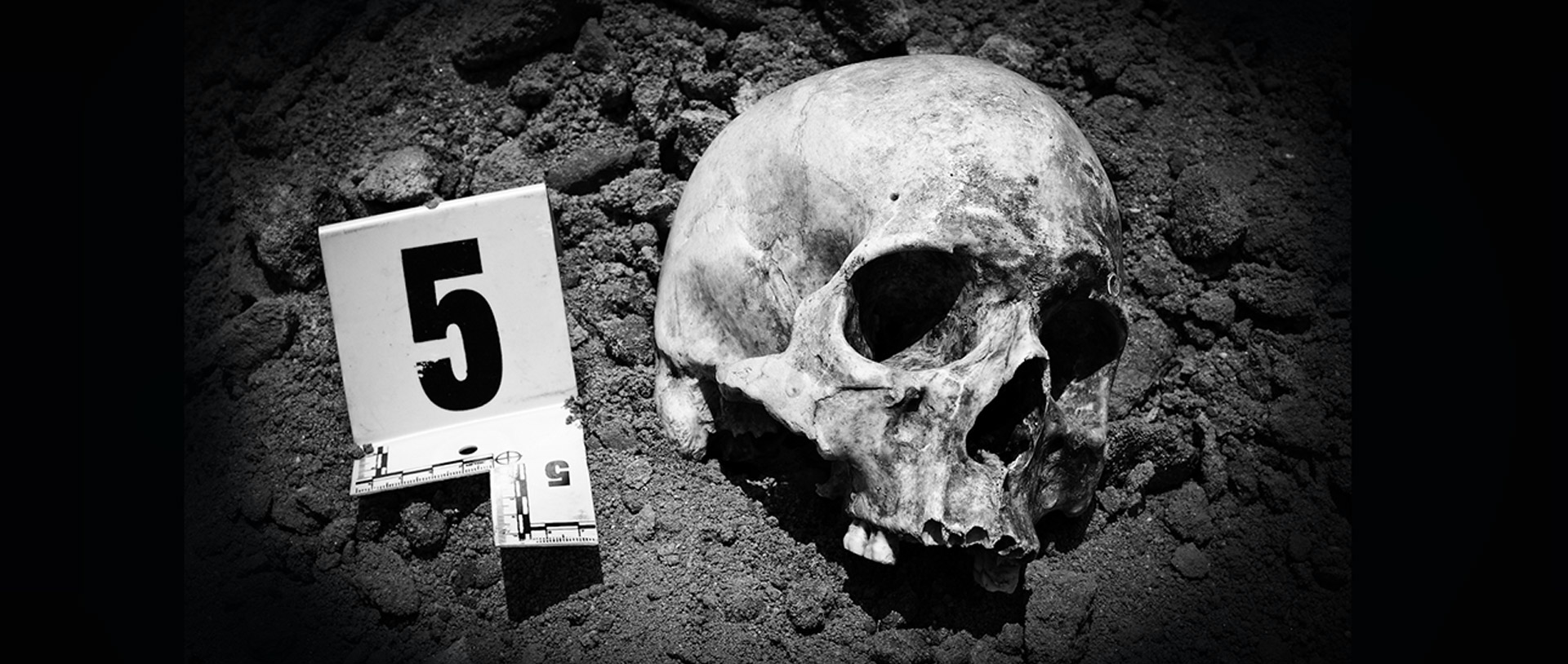
Conventionally, radiographs have been employed to record details such as remains, potential traumas, and distinctive features such as healing trauma or frontal sinus morphology. The evolution of advanced imaging technologies has significantly enhanced the ability to document skeletal remains and trauma in forensic anthropology cases.
Advancements in technology, particularly within the medical and engineering domains, have given rise to a variety of sophisticated imaging techniques, including computed tomography and Linear Slot X-Ray Technology. As these methods become more prevalent and their associated costs decrease, their integration into the field of forensic anthropology is increasingly common.
Harnessing Tech for Forensic Anthropology: Imaging Trends
Moving beyond the traditional use of photographs and radiographs, some forensic anthropologists employ virtual 3D models of skeletal material for purposes such as case analyses, documentation, and research. However, not all forensic anthropologists utilize these advanced technologies due primarily to limitations related to cost, access, and utilization.
Although technological advances in radiography have progressed in sophistication, quality, and cost of analysis (such as CT), digital radiography remains the most widely used imaging modality in forensic anthropology, second only to basic photography.
Forensic anthropology laboratories commonly feature some form of X-ray equipment, although the specific type may vary. Laboratories may possess closed table-type systems or open X-ray systems resembling those in hospital clinics.
In some cases, laboratories can boast a digital X-ray system that allows them to quickly take radiographs, enabling anthropologists to promptly assess the remains, selecting the parameters necessary for the study, such as Fobos XR, which can obtain high-resolution images of bones in less than 23 seconds.
Fobos XR: Empirical Evidence and Innovation in Forensic Anthropology
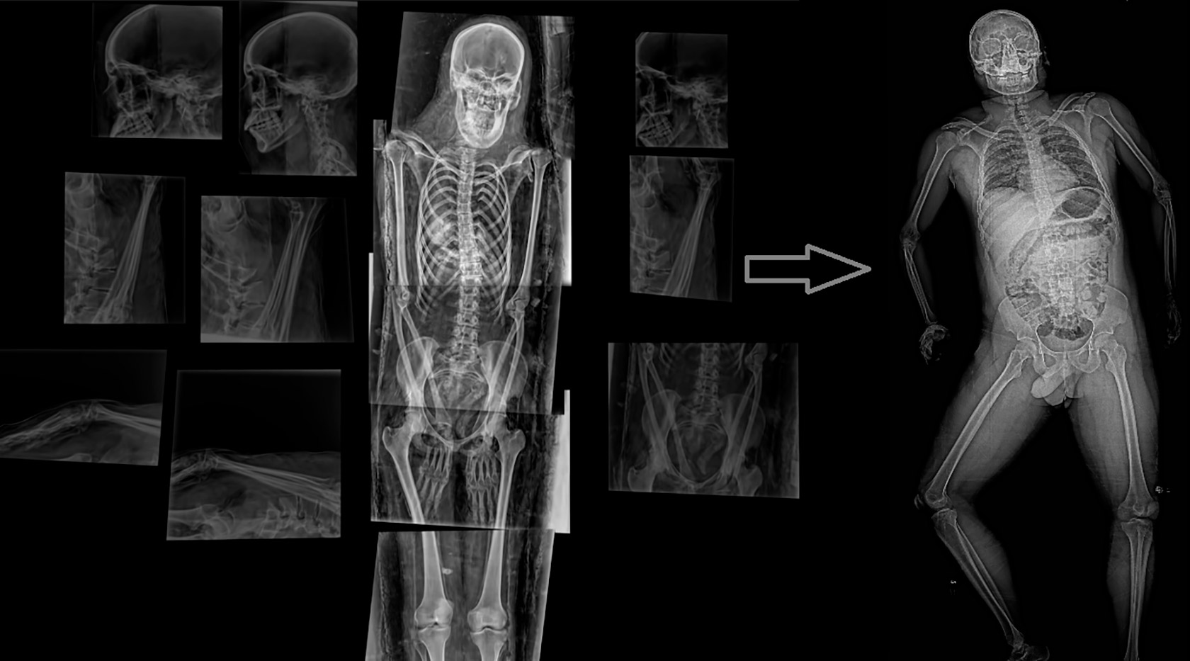
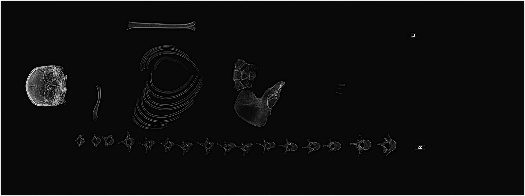
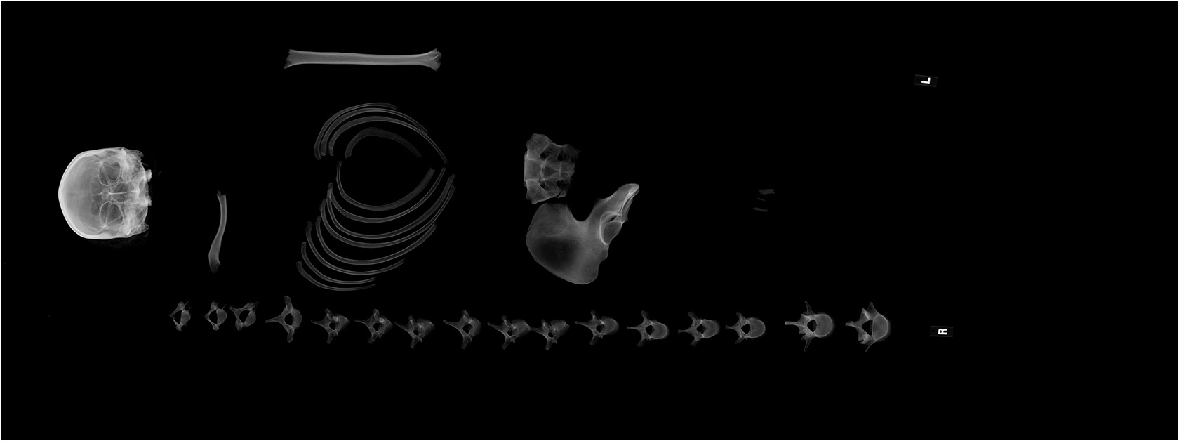
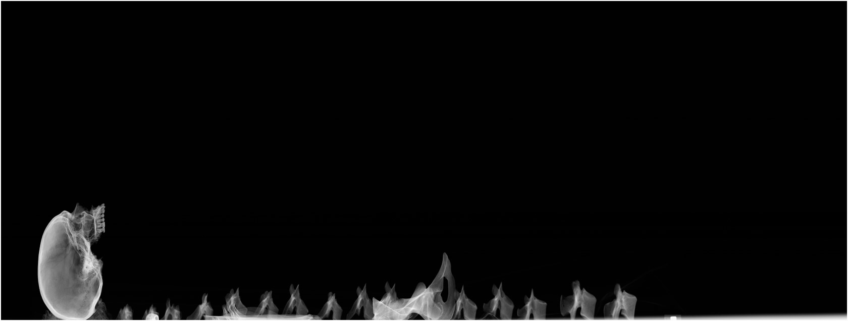
This unique technology provides a comprehensive view of bones from any angle (0 to 110 degrees). The device features a unique deck capable of lifting up to 295 kg for different types of bones. Its distinctiveness lies in a broad scanning field (207 cm), allowing a single scan to capture a full-large field image without the need to stitch multiple images together.
Recent innovations include partial scanning, offering visualization within a user-specified angle range (e.g., 0-90 degrees) with subsequent in-depth analysis. In 2021, research conducted on the Fobos XR machine focused on remains to identify gender and age from two perspectives, which are elaborated upon in this article.
Equipped with a high-quality CCD sensor, it captures high-resolution images with minimal noise, facilitating the easy diagnosis of clear bone structures. The scanner provides images with a pixel size of 100 µm and a spatial resolution of up to 5 lp/mm, enabling the identification of minute damages, deviations, and pathologies on bones.
Innovations in Radiography: The Fobos XR Advantage
To cater to the evolving needs of anthropologists, Linev Systems presents the innovative mobile forensic radiography system – FOBOS. This system reduces time and costs associated with transporting artifacts to stationary laboratories, ensuring the safety of valuable discoveries and minimizing the risk of damage or loss during transportation.
In many ways, forensic anthropologists employ radiography similarly to forensic pathologists. In instances involving suspected gunshot wounds, radiographs serve not only to identify potential bullets or fragments but also to detect areas of lead transfer.
Notably, even in fully skeletonized crania exposed to the elements for extended periods, radiographic evidence of lead around entrance and exit wounds or on impacted endocranial surfaces is observable. Thus, documenting lead transfer aids forensic anthropologists in deciphering bullet trajectories.


When considering the capabilities of the FOBOS XR System, its ability to capture images from various angles empowers anthropologists to trace the trajectory of a bullet comprehensively. This feature enables in-depth analysis, complemented by visual evidence. Real-life examples illustrated below showcase the system’s effectiveness in this regard.
Radiographs are crucial for investigating and documenting various forms of trauma, including fracture patterns. It should be standard operating procedure (SOP) in every forensic anthropology laboratory to radiograph all received remains, irrespective of their condition.
Cases vary, and not all received remains are completely skeletonized; some may have mummified or decomposing tissue. Radiographing all remains upon receipt allows the forensic anthropologist to demonstrate that fractures or skeletal trauma existed and were documented before any processing, providing empirical evidence that observed trauma is not a result of subsequent maceration or processing procedures.
Additionally, radiographing large masses of decomposing tissue ensures the confirmation of recovering all skeletal elements or potential evidence. Even when remains are fully skeletonized, radiographs of all elements remain essential. Furthermore, radiographs may unveil valuable characteristics within internal bone structures. Epiphyseal lines of fusion, visible radiographically, offer insights into the individual’s age.
Radiographs can also reveal signs of bony remodeling or disruptions in trabecular bone patterns, potentially indicating antemortem trauma, valuable in identifying the decedent or recognizing signs of potential abuse. Forensic anthropologists have employed radiographs to facilitate positive identifications, drawing a parallel to the way forensic odontologists use dental radiographs for comparisons between antemortem and postmortem conditions.
Similarly, forensic anthropologists can conduct radiographic comparisons across various skeletal regions. Numerous studies have demonstrated the uniqueness of frontal sinus morphology, observable through radiography, making it a reliable tool for forensic identifications.
Other radiographic studies have utilized distinctive bone shapes and features, suture patterns, or trabecular bone patterns to aid in identification. This approach proves particularly valuable in mass disaster scenarios, where antemortem information can be extracted from a finite pool of potential victims.
Skeletal variations in any body region may contribute to presumptive or positive identification, underscoring the importance of providing forensic anthropologists with antemortem radiographs alongside skeletal remains for the evaluation of unique morphologies.


Conclusion
In the past decade, the utilization of advanced imaging in forensic research and case analyses has experienced significant growth. However, these technologies have not yet become standard practice, and their full potential remains untapped in the field of forensic anthropology.
A major limiting factor is the cost, with computed tomography scanners and high-end laser scanners likely exceeding the budget constraints of forensic anthropologists. Here the FOBOS XR technology comes to the rescue, which has a not so high cost, but at the same time almost completely covers all the needs of anthropologists.